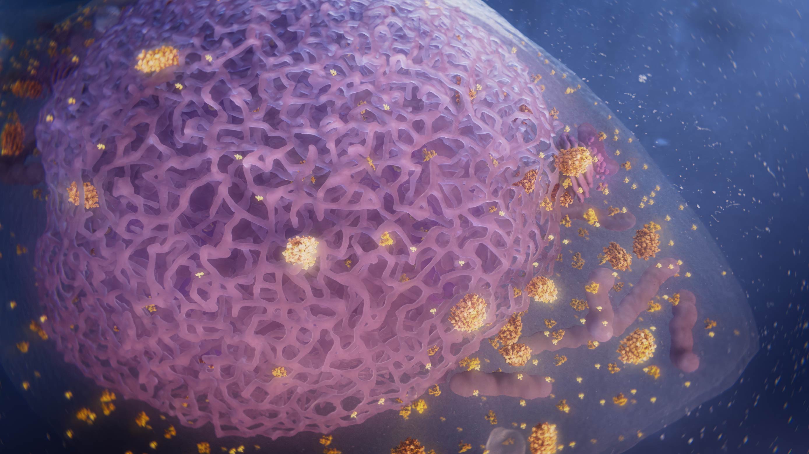
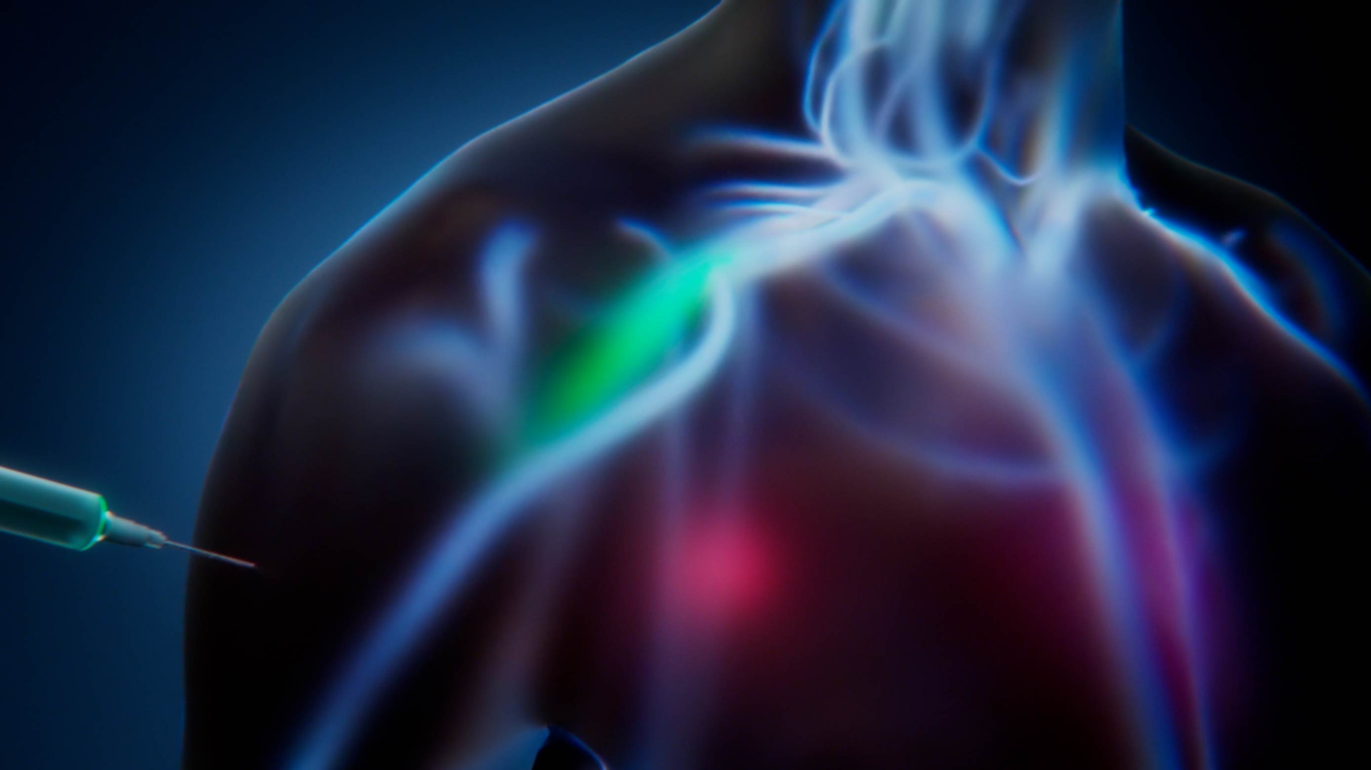
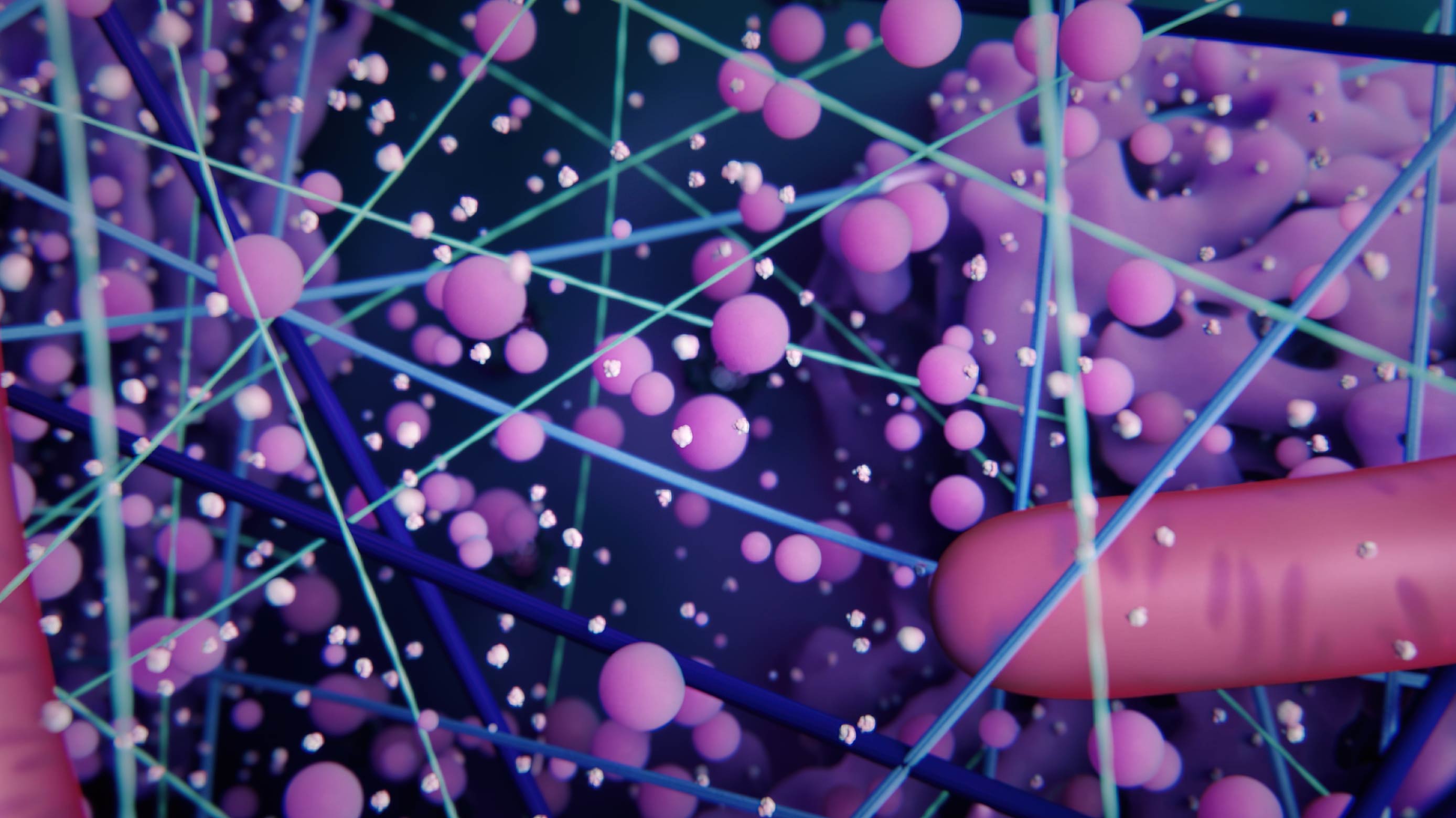
Mechanism of Action of the ENT-01 Drug


Project Goal
Create an explainer animation for investor relations and communications covering several aspects of Parkinson's disease and illustrating the mechanism of action of our client's ENT-01 drug.
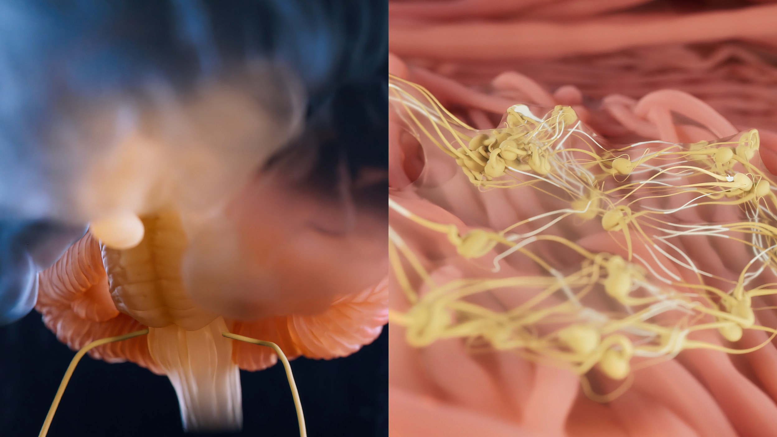
Brainstem anatomy and enteric neurons

Neurotransmitters in the normal synapse
Nerve signals along the gut-brain axis

ENT-01 displacing the alpha-synuclein aggregates
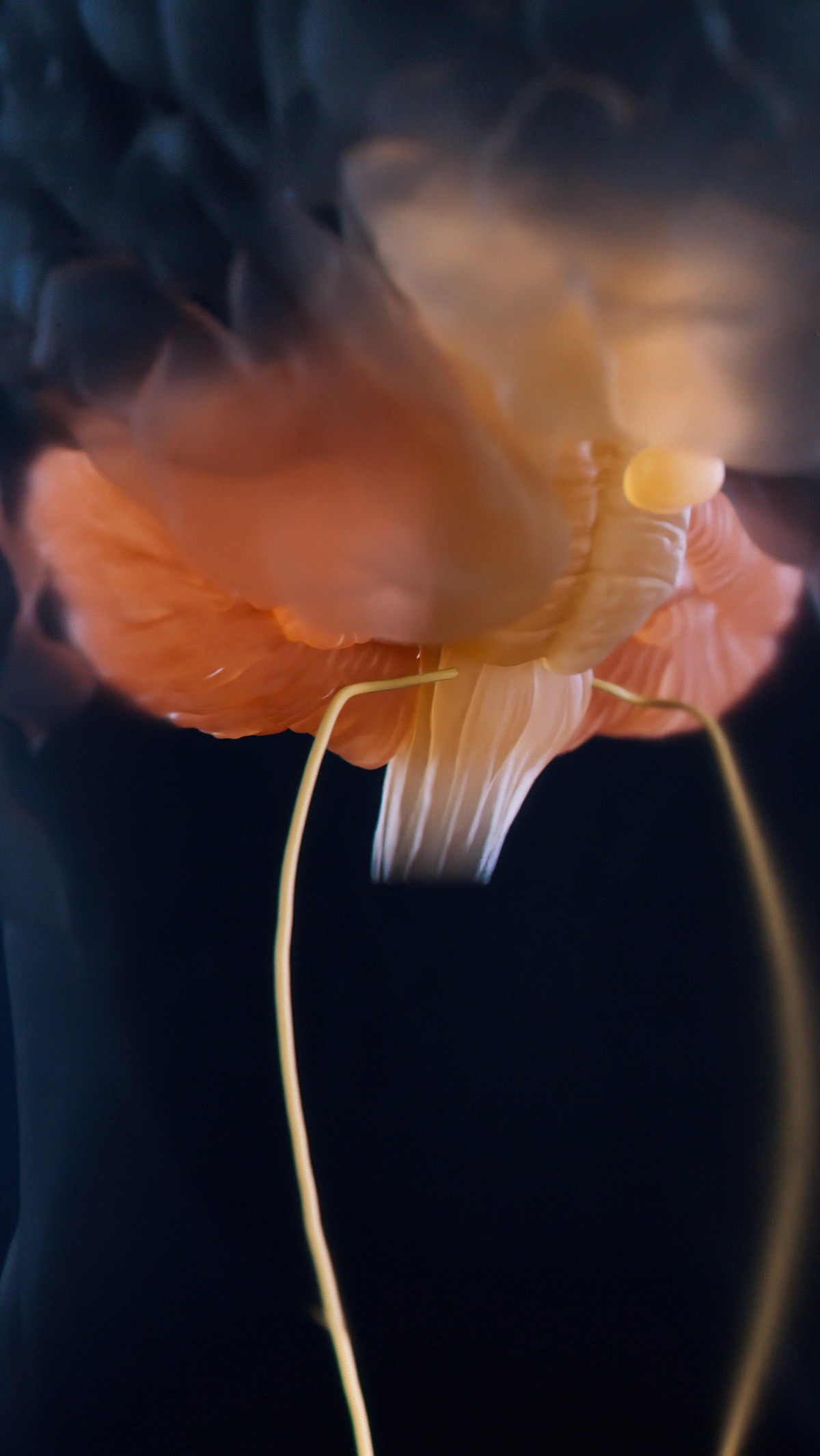
Visualization of brainstem and vagus nerve
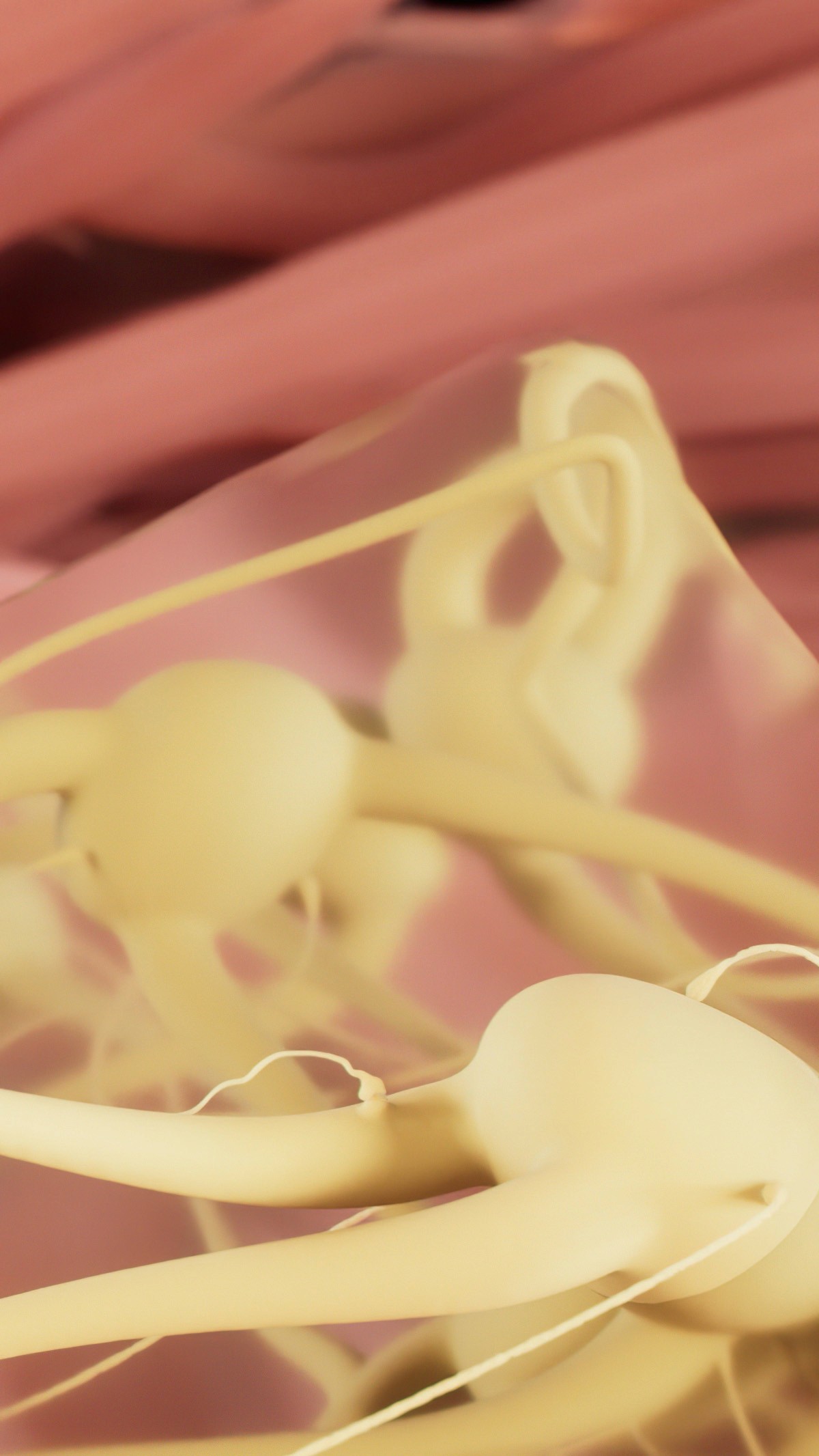
Enteric neurons in the submucosal plexus
The Process
Animation type:
3D + Deck 3D
Project timline:
10 weeks
Review:
5 zoom calls + 5 e-mail feedback sessions
Find out which of our 26 scientific animation options works best for investor relations and communications:
This project followed a well-tested workflow, consisting of seven production steps and four review stages. At each stage, we shared key updates on progress. Thanks to our deep expertise in both the scientific field and investor relations, clients typically have only minor suggestions to contribute, which was true for this project as well.
At every review stage, we implemented all minor edits based on client feedback and made the necessary adjustments. On average, a client dedicates about 5-9 hours to engover the entire project. We are flexible with communication, whether via email or Zoom calls, adapting to what suits our clients best.
Communication between the client and the Visual Science team is managed by a PhD-qualified account manager, ensuring our interactions are as smooth and efficient as possible, supporting medical communication services for complex scientific projects.
Why did we use this
animation type?
This animation was longer than our average project, meaning that the production was more resource-intensive. To balance our client's needs and budget, we offered a simplified 3D style, with fewer details in the cellular and molecular visualizaions. The result is still eye-catching and scientifically accurate. Simplified 3D also helps to direct the viewer's attention to the key aspects and messages without unnecessary details.
Despite the less detailed molecular and cellular models, our anatomical visualizations end up almost as detailed as regular 3D medical animations. The anatomical scenes required advanced 3D sculpting and animation of the peristalsis. Meanwhile, most of the models of the central nervous system were taken from the library of 3D assets which we have assembled over years of successful work. Some of the assets were reconstructed from CT scans.