Scientifically accurate 3D model and animation of HIV

Project Goal
Create the most scientifically accurate and detailed 3D model of the HIV particle in atomic resolution.

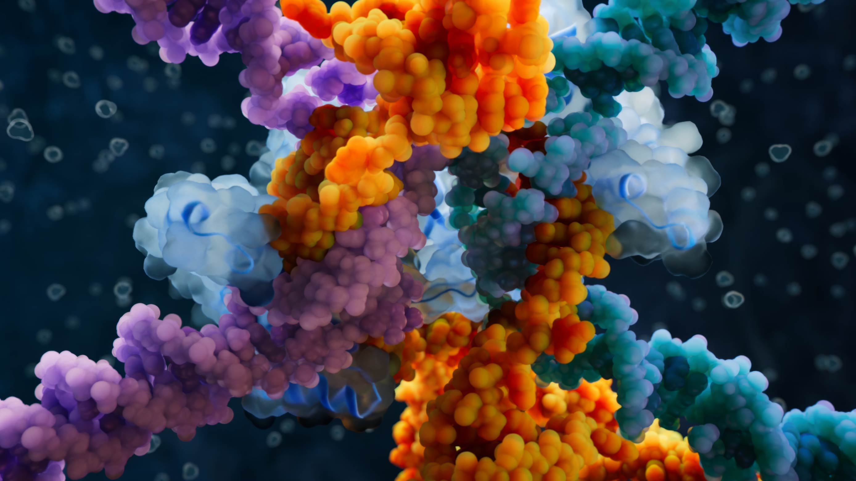
HIV whole model visualization

Surface proteins on the HIV particle
Envelope and capsid of the HIV particle


Nature Medicine special issue cover

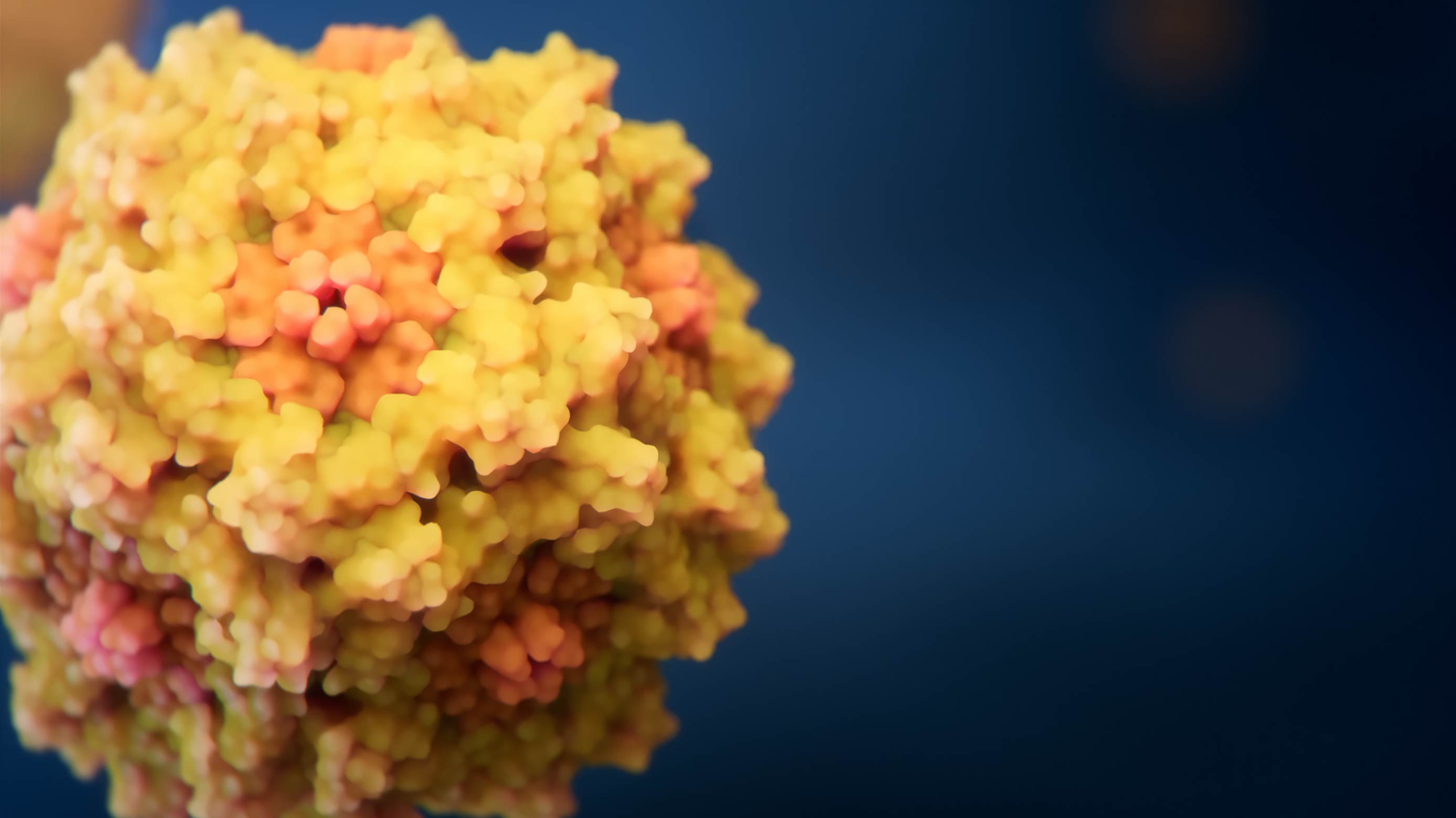
The particle core
Envelope proteins on the HIV surface
The Process
Animation type:
High-end 3D based on science grade molecular simulations
Project timline:
16 weeks
Find out which of our 7 scientific animation types and 20 subtypes work best for your project
This model summarizes results from more than 100 scientific publications published prior to 2009. Our team spent many weeks collecting and analyzing data, consulting with virologists, and assembling and refining the molecular structures of HIV components, reflecting scientific content development services grounded in primary literature. None of the currently available scientific methods allow for obtaining an image of the entire virus particle at atomic resolution.
Nevertheless, hundreds of studies by researchers around the world have shed light on the structure and morphology of virion components and their interactions, making the creation of this model possible.
The first-ever molecular animation based on science-grade molecular simulations:
This complex and highly resource-intensive project was the first high-end scientific animation and visualization project we produced, and it set standards not only for our team but also for many of our colleagues in the industry. The depicted spatial configurations of 17 different viral and cellular proteins present in the HIV particle are strictly consistent with known 3D structures.
The viral membrane in the model includes approximately 160,000 lipid molecules of eight different types, in proportions consistent with those found in the HIV particle.